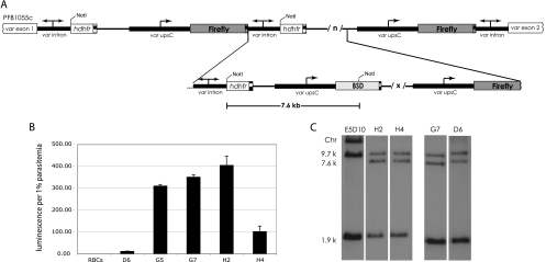
Fig. 3.
Multiple-copy pVBH integration. (A) Schematic of pVBH integration into the original pVLHIDH concatemer. Multiple copies (/x/) of pVBH integrated into the 3′ region of the original pVLHIDH concatemer. (B) Luciferase expression from uninfected cells (RBCs) and the clones D6, G5, G7, H2, and H4. The error bars indicate standard deviations. (C) Representative Southern blots showing four genotypically indistinguishable clones. gDNA digested with NotI and probed with hdhfr show a loss of the large chromosomal band (Chr), indicating integration after the last hdhfr in the original concatemer. The 7.6-kb band also hybridizes to a bsd probe (not shown).

