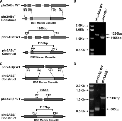
Fig. 1.
Generation of phr2aBα and phr2aBβ knockout cell lines and analysis via genomic PCR. (A) The top panel shows a schematic of gene replacement by homologous recombination, indicating the wild-type (WT) phr2aBα locus and the blasticidin-resistant knockout construct that was generated using PCR primers P1 to P4. Exons (boxes) and introns (dotted lines) are indicated. The vertical dotted lines indicate predicted homologous recombination regions. The bottom panel indicates primers used for the PCR screening of the knockout clones. (B) Agarose gel electrophoresis of the genomic PCR screening presenting profiles of parental and phr2aBα knockout products. (C) The top panel shows a schematic of gene replacement by homologous recombination, indicating the wild-type (WT) phr2aBβ locus and the blasticidin-resistant knockout construct that was generated using PCR primers P5 to P8. The vertical dotted lines indicate predicted homologous recombination regions. The bottom panel indicates primers used for the PCR screening of the knockout clones. (D) Agarose gel electrophoresis of the genomic PCR screening presenting profiles of parental and phr2aBβ knockout products.

