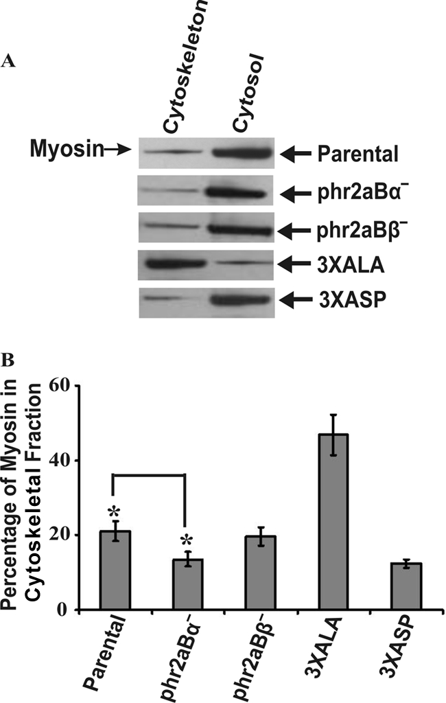
Fig. 5.

Quantitation of myosin II assembly in phr2aBα- and phr2aBβ-null cells into Triton-resistant cytoskeletal ghost fractions. (A) Shown are sample SDS-PAGE/Western blot profiles used for quantitative analysis of percentage of myosin II assembled in the Triton-resistant cytoskeletal fractions in parental, 3XALA, 3XASP, and phr2aBα- and phr2aBβ-null cell lines. (B) Quantification of myosin II present in Triton-insoluble fractions in each cell line, determined from densitometry of Western blots. Error bars represent the standard errors of the mean (SEM) with 10 independent samples. *, P ≤ 0.005.
