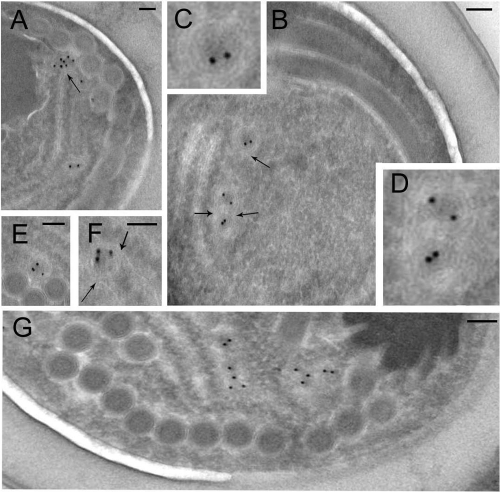
Fig. 2.
Identification of mitosomes in A. locustae spores. Ultrathin cryosections of mature spores were stained with affinity-purified and depleted anti-mitHsp70 Abs. Immunoelectron microscopy shows specific labeling of mitosomes, small (50- to 200-nm) structures of round profiles bounded by a double membrane (A to G). About 3 or 4 mitosomes were found per spore section, and some of them were aggregated in pairs (B, G). Mitosomes from panel B are shown enlarged (C, D). As expected, most of the labeling was present over the inner area of the organelles (mitosomal matrix). Microsporidial mitosomes appeared to be in close contact with ER cisternae (arrows). Gold grain size, 10 nm. Scale bar, 0.1 μm.

