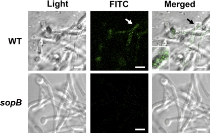
Fig. 3.
Translocation of SopB into C. albicans filaments via SipB. SopB translocation in S. Typhimurium wild-type (top row) and sipB mutant (bottom row) was visualized by confocal microscopy. Scale bars, 4.2 μm. The inset shows an enlarged view of the area indicated by the arrow. An immunofluorescence assay was performed as described in Materials and Methods.

