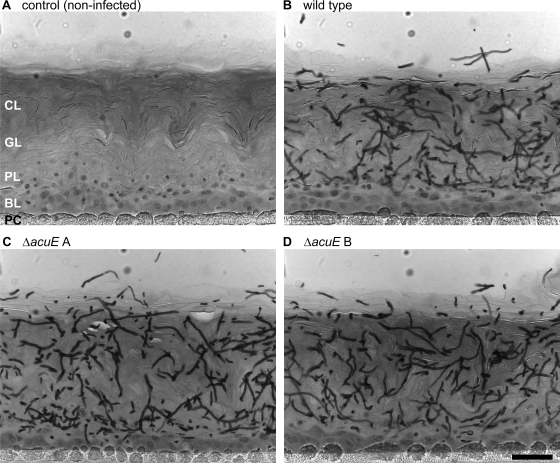
Fig. 9.
Comparable epidermal invasion of A. benhamiae wild-type and ΔacuE mutant cells in a model of RHE. The RHE was infected with 1 × 10³ A. benhamiae microconidia and incubated at 37°C in 5% CO2. Invasion of the RHE was analyzed 4 days after infection by microscopy as described in Materials and Methods. Microscopy of representative PAS-stained sections shows similar degrees of fungal invasion of the RHE by the wild-type strain Lau2354-2 (B) and the mutant strains AbenACUEM1A (C) and AbenACUEM1B (D). An uninfected control sample is shown for comparison (A), and the scale bar represents 50 μm. CL, cornified layer of the RHE; GL, granular layer; PL, prickle cell layer; BL, basal layer; PC, polycarbonate filter.

