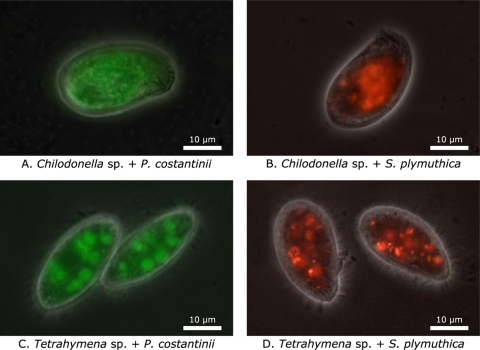
Fig. 2.
Representative Chilodonella sp. (A and B) and Tetrahymena sp. (C and D) cells after feeding on GFP-expressing P. costantinii (A and C) or RFP-expressing S. plymuthica (B and D) biofilms. Ciliates were added to bacterial biofilms 45 min before being fixed with formalin. Composite images constructed from phase-contrast microscopy images overlaid with fluorescence microscopy images are shown.

