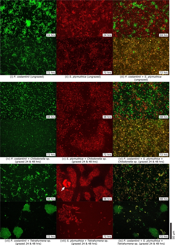
Fig. 4.
Representative confocal microscopy images of live bacterial biofilms. Ungrazed biofilms of P. costantinii (i), S. plymuthica (ii), and both bacteria together (iii) after 24 and 72 h of growth. Chilodonella sp.-grazed biofilms of P. costantinii (iv), S. plymuthica (v), and both bacteria (vi) after 48 and 72 h (24 h of biofilm growth followed by 24 h and 48 h of grazing). Tetrahymena sp.-grazed biofilms of P. costantinii (vii), S. plymuthica (viii), and both bacteria (ix) after 48 and 72 h (24 h of biofilm growth followed by 24 and 48 h of grazing). The S. plymuthica-filled vacuoles of a feeding Tetrahymena sp. cell are indicated (arrow). Green fluorescence indicates P. costantinii cells. Red fluorescence indicates S. plymuthica cells. Yellow fluorescence in biofilms composed of both bacteria also represents S. plymuthica cells and is the result of superimposed red and green fluorescence due to the use of high-intensity blue laser illumination causing simultaneous fluorescence of both S. plymuthica and P. costantinii cells in combination with a dual-wavelength filter. The scale bar applies to all images.

