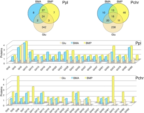
Fig. 1.
Distribution of P. placenta (Ppl) and P. chrysosporium (Pchr) proteins identified by LC-MS/MS in BMA and BMP culture filtrates after 5 days of growth. Upper Venn diagrams show the partitioning of glucose-, BMA-, and BMP-derived proteins. In the lower panels, the numbers of proteins identified within glycoside hydrolase families are illustrated.

