Figure 4.
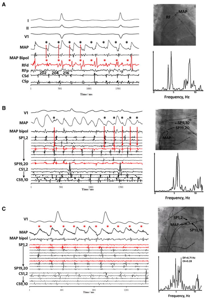
Complex fractionated electrogram (CFAE) caused by far-field signals (dissociated from local monophasic action potential [MAP]). A: CFAE at left atrial appendage base. MAPs adjacent to CFAEs are distinct, reflecting local activation and refractory period (action potential duration 150–170 ms). Superimposed signals (asterisks) are dissociated from local activation (MAP), may occur in very early repolarization, and thus appear far field. These signals coincide with RFd (distal RF) signals (arrows). B: CFAE at the right atrial appendage orifice. MAPs are again distorted by dissociated electrograms (asterisks) throughout repolarization that likely are far field, coinciding with signals on SP9,10 and SP19,20 (red) of the spiral catheter. C: CFAE near left superior pulmonary vein. Intermittent CFAE timed with small electrograms (asterisks) dissociated from local MAP and electrograms on nearby SP1,2 and SP13,14 (red) spiral electrodes. In each panel, spectral dominant frequency is broad.

