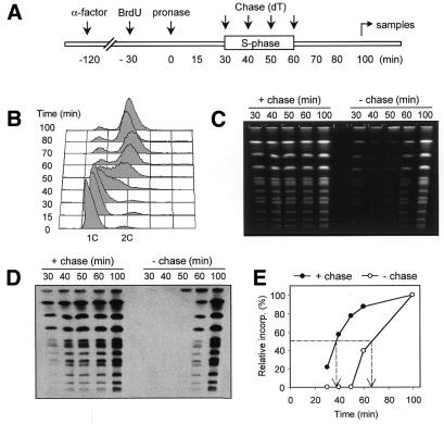
Figure 4.
Pulse–chase incorporation of BrdU during S phase. (A) TK+ cells (E1000) arrested in G1 with α-factor for 2 h were released by addition of 50 µg/ml pronase in medium containing 200 µg/ml BrdU at 25°C. The culture was split in two and cells were either collected at 10 min intervals during the following S phase (–chase) or were chased with a 10-fold excess of thymidine (dT) at the same time points (+chase). In the latter case, cells were only harvested in G2 (100 min). (B) FACS profile of the pulse–chase experiment. (C) PFGE analysis of the +chase and –chase experiments. The electrophoresis was performed as described in Materials and Methods and the gel was stained with ethidium bromide. (D) Detection of BrdU incorporation. Southern blotting and detection were performed as described in Figure 1, except that a secondary antibody coupled to HRP was used and revealed with an ECL reaction (Amersham). (E) Quantification of BrdU signals on the Southern blot shown in (D). As only fully replicated chromosomes enter the gel in the –chase experiment, the shift on the x-axis between the two curves indicates S phase duration.

