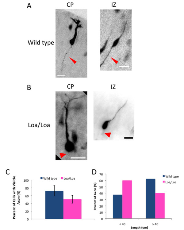
Figure 7.
Loa/Loa neurons display a defect in axon extension in vivo. (A,B) Individual neurons from Oregon Green stained wild-type (A) or Loa/Loa (B) brain sections with a visible axon (thin arrowhead), or no visible axon (wide arrowhead). Neurons from both the cortical plate (CP) and intermediate zone (IZ) were analyzed. Scale bars = 20 μm. (C) Quantification of the percentage of wild-type or Loa/Loa neurons with a visible axon. Only neurons with visible leading processes were scored for an axon, or lack thereof. Error bars represent the standard deviation. (D) Quantification of the percentage of wild-type and Loa/Loa axons that were shorter or longer than 40 μm.

