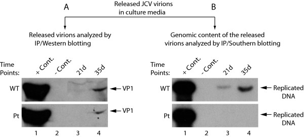
Figure 7.
Analysis of the virions released from PHFG cells transfected/infected with either JCV Mad-1 WT or its Pt mutant. Supernatants from the transfected/infected PHFG cells were collected at indicated time points centrifuged at 16,000 × g to clear the cell debris and were subjected to immunoprecipitation using an anti-VP1 antibody (PAb597) to precipitate the released virions. Immunoprecipitants were divided into two equal portions, one of which was analyzed by Western blotting to detect viral capsid protein VP1 (A) and the other was analyzed by Southern blotting for the detection of the encapsidated viral DNA (B). In lane 1 in panel A, supernatant from infected cells was immunoprecipitated with anti-VP1 antibody and loaded as a positive control (+ Cont.). In lane 2 in panel A, supernatant from uninfected cells was immunoprecipitated with anti-VP1 antibody and loaded as a negative control (- Cont.). In lane 1 in panel B, JCV Mad-1 genome (2 ng) digested with BamH I was loaded as a positive control (+ Cont.). In lane 2 in panel B, supernatant from uninfected cells was immunoprecipitated, digested with BamH I and loaded as a negative (- Cont.).

