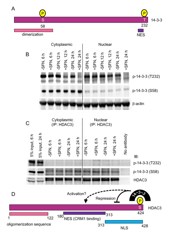
Figure 8.
Role of 14-3-3 phosphorylation status in HDAC3 binding. (A) Domains in 14-3-3 showing phosphorylation sites probed by immunoblotting. (B) Nuclear and cytoplasmic extracts from HCT116 cells treated with 15 μM SFN or DMSO were immunoblotted with phosphospecific antibodies to p-14-3-3(T323) and p-14-3-3(S58). (C) HDAC3 pull-downs, performed as in Figure 7, were followed by immunoblotting for p-14-3-3(T323), p-14-3-3(S58), and HDAC3. (D) Model for 14-3-3 interacting with HDAC3: repressive actions on the nuclear localization signal (NLS) in 14-3-3, plus possible activation of the nuclear export signal (NES), are proposed based on prior studies with class IIa HDACs [56].

