Figure 1.
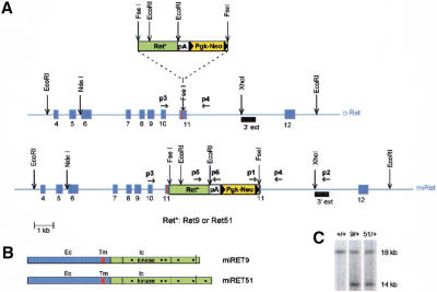
Targeting of the c-Ret locus. (A) Schematic representation of the targeting strategy. A cassette (top) consisting of cDNA fragments encoding the intracellular part of human RET9 or RET51 (Ret*), the β-globin polyadenylation signal (pA), and the NeoR gene (Pgk-Neo) was inserted in frame into exon 11 of the mouse locus, immediately after the segment encoding the transmembrane domain (red stripe, middle). The targeted locus is shown at the bottom. Solid arrowheads indicate loxP sites. (B) The targeted loci encode single chimeric RET receptors with identical extracellular (Ec) and transmembrane (Tm) domains and the intracellular (Ic) segment of human RET9 or RET51. The positions of Tyr-residues thought to be important for RET signaling are indicated by dots. (C) Southern blot analysis of DNA from ES cell clones using the probe shown in A (3′ ext). On digestion with EcoRI, the wild-type and targeted loci generate 18-kb and 14-kb fragments, respectively.

