Figure 3.
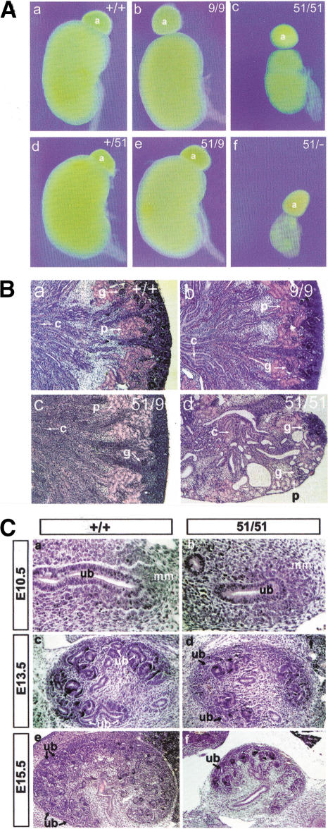
Renal hypodysplasia in miRet51animals. (A) Kidneys from miRet9/miRet9 (b), +/miRet51 (d) and miRet9/miRet51 (e) animals appeared identical to the wild type (a). The kidneys of miRet51/miRet51 (c) or miRet51/Ret.k− (f) animals, however, were hypodysplastic. +/+, n = 134; 9/9, n = 43; 51/51, n = 103; +/51, n = 197; 9/51, n = 35; 51/−, n = 17. a, Adrenal gland. (B) Histological analysis of kidneys of miRet neonates. The histoarchitecture of miRet9/miRet9 (b) and miRet9/miRet51 (c) kidneys was indistinguishable from the wild type (a). However, the kidneys of miRet51/miRet51 neonates (d) show many cysts and a reduced number of nephrons. g, Glomerulus; p, proximal convoluting tubule; c, collecting tubule. For all genotypes n = 7. (C) Reduced branching of the UB in miRet51 homozygote embryos. Sections through the metanephros of E10.5 +/+ (a) and miRet51 (b) embryos reveal no difference in the evagination of the UB (ub) and the invasion of the metanephric mesenchyme (mm). Note the normal condensation of the mesenchyme in the miRet51embryo. At E13.5, a small reduction in the number of UB branches is detected in the miRet51 embryos (d) in comparison with +/+ (c). This difference becomes clearer at subsequent developmental stages (E15.5; cf. panels e and f). For all genotypes and stages n = 5.

