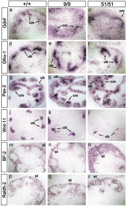
Figure 4.
Molecular analysis of metanephric development in miRet mice. In situ hybridization with riboprobes for Gdnf (a–c); Gfrα1 (d–,f); Pax2 (g–i); Wnt11 (j–l); BF-2 (m–o); and Raldh2 (p–r) on serial sections of E13.5 kidneys from +/+ (a,d,g,j,m,p), miRet9 (b,e,h,k,n,q), and miRet51 (c,f,i,l,o,r) embryos. cm, Condensing mesenchyme; ub, ureteric bud tips; st, stroma. For each genotype and probe n = 4.

