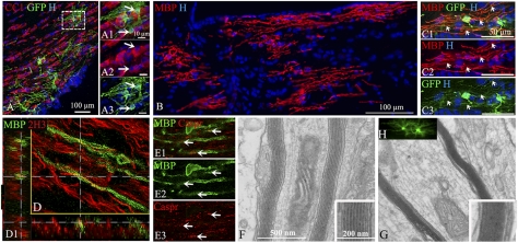
Fig. 4.
Differentiation of BC progeny into myelin-forming oligodendrocytes after long-term transplantation. (A–A3) GFP+ cells (green; A and A2) give rise to mature CC1+ oligodendrocytes (red; A1 and A3) in corpus callosum. (B) Immunolabeling for MBP indicates widespread BC-derived MBP+ myelin patches in the fimbria. (C1–C3) Illustration of GFP+ cells (green; C1 and C3) expressing MBP (red; C1 and C2) with features of myelin-forming oligodendrocytes extending processes to internodes (arrows). (D and D1) BC-derived MBP+ myelin segments (green) surround 2H3 host axons (red); D1 orthogonal views. (E1–E3) MBP+ myelin internodes (green; E1 and E2) express normally organized Caspr paranodal protein (red; E1 and E3). (F and G) Ultrastructural analysis confirms the presence of compacted donor-derived myelin (G Inset) compared with uncompacted shiverer myelin (F Inset). (H) Illustration of GFP+ oligodendrocytes in corresponding floating sections prior to processing for electron microscopy.

