Fig. 1.
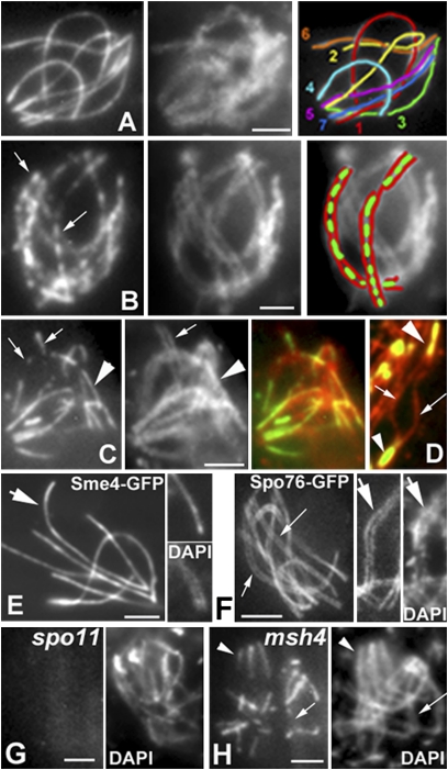
Sme4-GFP localization during meiotic prophase. (A–F) WT. (A) Pachytene. (Left) Sme4-GFP. (Center) DAPI. (Right) Drawing of the seven bivalents. (B) Early zygotene. (Left) Short Sme4 lines (arrows) along all bivalents. (Center) DAPI. (Right) Drawing of two bivalents (green, Sme4; red, DAPI). (C) Midzygotene. (Left) Mixture of short (arrows) and longer Sme4 lines (arrowhead). Correlated differences in pairing seen in DAPI (Center) and merge (Right). (D) Sme4 localization in relation to Rec8-RFP axis staining (red). Sme4-GFP is only seen in synapsed regions (arrowheads) and not in nonsynapsed region (arrows). (E and F) Sme4 and Spo76 localization at pachytene. Sme4 forms single bright lines (E Left, arrow) between the homolog chromatin (E Upper Right compared with DAPI in E Lower Right). Spo76-GFP illuminates double lines of chromosome axes (F Left and Center, arrows) corresponding to homolog chromatin (DAPI in F Right). (G Left) No Sme4-GFP staining in spo11 (DAPI in Right). (H Left) In the absence of Msh4, Sme4 is seen where homologs synapse (arrowheads) and not in synapsing regions (arrow; DAPI in Right). (Scale bars: 2 μm.)

