Fig. 6.
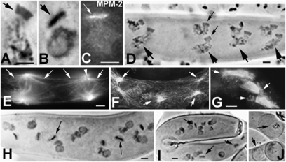
Postmeiosis in WT and mutants. (A, B, D, and H–J) Hematoxylin staining. (A–C) Frontal views of SPBs at PMM telophase. (A) WT SPB with typical rectangle shape. (B) sme4 SPB is flat and dense and is confirmed by MPM-2 staining (C). (D) WT Podospora ascus with the eight post-PMM nuclei arranged in four rows of two nuclei (large arrows); small arrows show one pair of SPBs. (E–G) Antitubulin staining. (E) Two WT PMM anaphase spindles. Large arrays of astral MTs (arrows) delimit clear areas along the ascus; arrowhead points to SPB (unstained rectangle). (F) Same stage as E in sme4. Flat SPBs nucleate only short astral MTs (arrows) that are intermingled with persisting cortical MTs (absent in WT). (G) pame4 spindles grouped in the center of the ascus because of defective astral MT-mediated (arrows) nuclear movement. In pame4 (H) and sme4 (I), post-PMM nuclei are arranged randomly and often degenerate (compare with WT chromatin in D) before spore enclosure. (J) Abortive ascospores (arrows). (Scale bars: 5 μm.)

