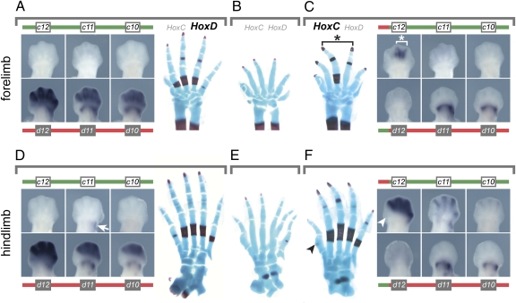
Fig. 3.
Hoxc genes can functionally substitute for Hoxd genes during digit development. (A) In E12.5 wild-type embryos, posterior Hoxd genes are expressed in both the proximal and the distal part of the growing forelimb. No transcript is detected for Hoxc12, Hoxc11, or Hoxc10. (B) In newborns, the absence of Hoxd gene function in digits leads to a reduction in size and malformation of the cartilage rods, as well as a delay in their ossification. (C) After translocation, Hoxd gene expression is lost distally, whereas Hoxc12 becomes expressed in digits III and IV, i.e., precisely where a rescue in both the size and the ossification pattern is observed (asterisk). (D) As for the forelimb, only Hoxd genes are expressed in the distal domain of the developing hind limb. Proximally, however, transcripts for Hoxc11 are detected (arrow). (E) The loss of distal Hoxd function induces similar effects on foot development, as observed in forelimbs. (F) In distal hind limbs at E12.5, Hoxc12, Hoxc11, and Hoxc10 are expressed in a quantitative collinear manner when controlled by the HoxD regulatory landscape, much like Hoxd genes in their wild-type context. These Hoxc transcripts substantially rescue the cortical ossification pattern in the foot, which is now almost identical to wild type. The big toe is not rescued, as explained by the lack of transcription of Hoxc genes in this presumptive digit (arrowheads).

