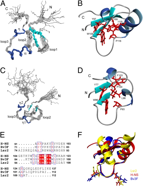
Fig. 3.
Solution structure of H-NSCtd and Bv3FCtd. Superimposition of backbone traces for the ensemble of 20 structures of H-NSCtd (A) and Bv3FCtd (C). Ribbon representation of H-NSCtd (B) and Bv3FCtd (D) mean structures. Only well-structured regions are included. Side chains of residues that form the hydrophobic core are shown in red. (E) Sequence alignment of the C-terminal domains of H-NS, Bv3F and Lsr2. (F) Superimposing structures of H-NSCtd (red), Bv3FCtd (blue), and Lsr2Ctd (yellow). The loop consisting of the conserved “T/SXQ/RGRXPA” motif adopts nearly identical conformation in these three structures.

