
Table 1.
Functionalization of Pcl proteins
| Reagent | Structure(s) | Coupling [%] | Fig. |
| PEGs (linear and branched) | 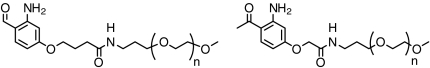 |
50 to > 95 | 3, 5 |
| Disaccharide | 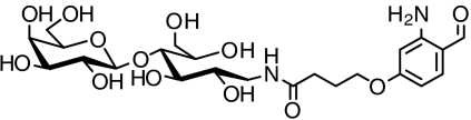 |
> 98 | SI Appendix: Fig. S8 |
| DOPE phospholipid | 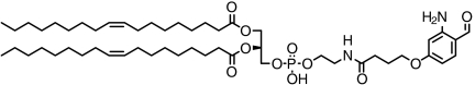 |
> 94 | SI Appendix: Fig. S9 |
| Biotin | 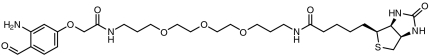 |
> 98 | SI Appendix: Fig. S10 |
| Fluorescein | 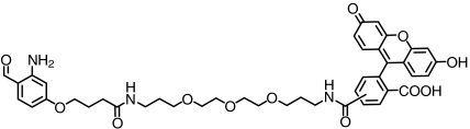 |
> 88 | SI Appendix: Fig. S11 |
| Nitro-phenyl haptens | 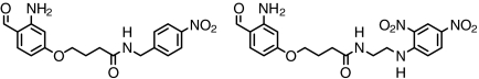 |
> 93 | SI Appendix: Fig. S12 |
| CpG- phosphothioate | 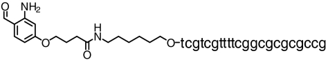 |
∼80 | SI Appendix: Fig. S13 |
| PADRE peptide (T-cell epitope) | 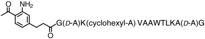 |
> 83 | SI Appendix: Fig. S14 |
Pcl reagents tested and coupling efficiencies at pH 7.4 and 22 °C. For details and results, see Fig. 3, Fig. 5, and SI Appendix: Figs. S8–S14 as indicated