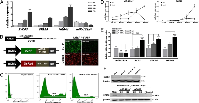
Fig. 5.
In vitro target assay of miR-181a* on the NR6A1 transcript and a quantitative expression analysis of meiotic genes following the knockdown of miR-181a* in chicken PGCs. (A) Expression levels of the meiotic genes, including SYCP3, STRA8, and NR6A1, were analyzed in chicken PGCs 0–72 h after RA treatment. Expression of endogenous miR-181a* in PGCs was down-regulated after RA treatment. (B) Diagram of the miR-181a* binding site in the NR6A1 3′ UTR and the expression vector maps for eGFP with NR6A1 3′ UTR and DsRed with miR-181a*. After cotransfection of pcDNA-eGFP-3′-UTR for the NR6A1 transcript and pcDNA-DsRed-miRNA for miR-181a*, the fluorescence signals of GFP and DsRed were detected using fluorescence microscopy and FACS (C). (D) Quantitative expression pattern of miR181a* and NR6A1 in male and female chicken embryonic gonads from E9.5 to E17.5. Error bars indicate the standard error (SE) of triplicate analysis. (E) Relative expression of SYCP3, STRA8, and NR6A1 after silencing with the knockdown probes for miR-181a* at 72 h, followed by RA treatment in chicken PGCs. *P < 0.05, **P < 0.01, and ***P < 0.001: significant difference compared with control. (F) Expression of SYCP3 protein after silencing with the knockdown probes for miR-181a* at 72 h, followed by RA treatment in chicken PGCs examined by Western blotting. OE, overexpression, KD, knockdown.

