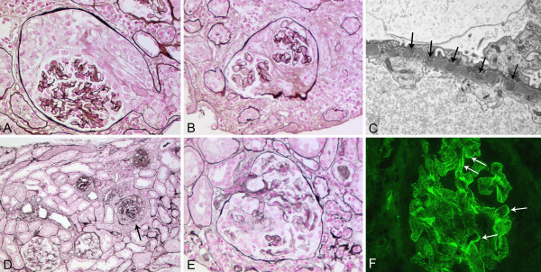
Figure 1.
Renal biopsy findings. The first biopsy (corresponding to month 0 in table 1) showed cellular crescents (A) in two and fibrocellular crescents (B) in three out of nine glomeruli, consistent with crescentic glomerulonephritis with moderate activity and moderate chronicity (Silver methenamine stain; original magnification, (A) ×200, (B) ×180). The glomerular basement membrane revealed multiple small holes, no spikes. Immunofluorescence study results were negative but on electron microscopy multiple small subepithelial deposits (arrows) indicative of membranous nephropathy stage 1 were visible (C, original magnification, ×10.500). In the second biopsy (corresponding to month 23 in table 1) widespread global sclerosis was present with sclerosis of 15 of 32 glomeruli and crescents in 5 glomeruli: two segmental fibrous crescents, two fibrocellular (one gobal (arrow in D)), one segmental cellular crescent, with focal interstitial fibrosis and tubular atrophy, comprising 25% of the cortex, consistent with late ANCA-associated sclerosing glomerulonephritis with minimal activity and severe chronicity (Silver methenamine stain; original magnification, (D) ×50, (E) ×160). Immunofluorescence studies showed finely granular glomerular basement membrane positivity for IgG (2+) corresponding to the previously diagnosed membranous nephropathy and a segmental linear (arrows) GBM positivity (F original magnification, ×280).

