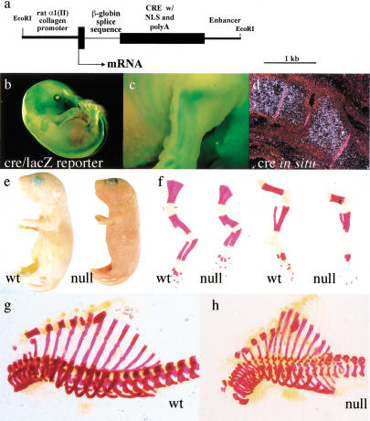
Figure 2.
Generation of mutant animals lacking HIF-1α in growth plate chondrocytes. (a) Schematic representation of the colIICre transgene. (b,c) Whole-mount β-galactosidase staining of E15.5 embryo. (d) in situ hybridization analysis with antisense Cre riboprobe on histological sections of wild-type hindlimb from E15.5 embryo. (e) Picture of newborn wild-type ( left) and null mouse littermates (right). (f–h) Whole skeleton Alizarin Red S staining; newborn wild-type (left) and mutant (right) forelimbs (left) and hindlimbs (right) are shown in f; newborn wild-type rib cage is shown in g; newborn null rib cage is shown in h.

