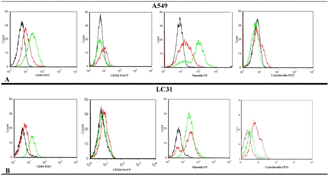
Figure 2. TGF-β1 up-regulates mesenchymal markers expression and down-regulates epithelial markers expression.
A: Cytometric analysis for CD90, CD326, vimentin and Cytokeratin in untreated (line red) and TGFβ-1 treated [line green] in A549 cell line after 20 days of treatment; B: Cytometric analysis for CD90, CD326, vimentin and Cytokeratin in untreated [line red] and TGFβ-1 treated (line green) in LC31 cell line after 30 days of treatment. Isotype controls are in black.

