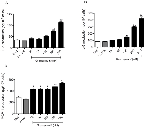
Figure 2. GrK stimulates IL-6, IL-8 and MCP-1 protein production in lung fibroblasts.
(A–C) HFLs were exposed to concentrations of GrK ranging from 10–300 nM at for 24 h. Cell culture supernatants were then collected and analyzed for IL-6, IL-8 and MCP-1 production by ELISA. Cell counts were used to normalize cytokine concentrations by controlling for variability in cell numbers. Control wells include cells treated with media alone or cells treated with heat in-activated GrK (100 nM). All data are expressed as mean protein production (pg/106 cells) ± SEM from three independent triplicate experiments. * p<0.05 when compared to media alone; ** p<0.01 when compared to media alone.

