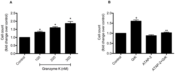
Figure 7. GrK induces cell proliferation in HFLs in a dose-dependent manner.
(A) Cells were incubated with 100 and 200 nM of GrK for 48 h then trypsinized and counted using a hemocytometer to determine cell number. (B) PAR-1 neutralization with ATAP-2 (5 µg/ml) abolished GrK (200 nM)-induced cell proliferation. Data are expressed as mean fold change ± SEM from three independent experiments run in triplicate. * p<0.05 compared to media control.

