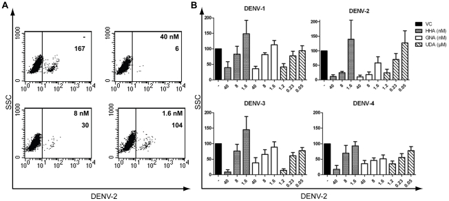
Figure 2. Dose-dependent antiviral activity of HHA, GNA and UDA in DENV-infected MDDC.
(A) MDDC were infected with DENV-2 in the absence (−) or presence of dose-dependent concentrations of HHA. The number of DENV-2 positive cells was determined by flow cytometry using 5 µg/ml anti-DENV antibody recognizing the E-protein of DENV-2 (clone 3H5). In each plot, the number of DENV positive cells is indicated. (B) MDDC were infected with the four serotypes of DENV in the presence or absence of various concentrations of HHA, GNA and UDA. DENV infection was analyzed by flow cytometry using an anti-PrM antibody recognizing all four DENV serotypes (clone 2H2). % of infected cells compared to the positive virus control (VC) ± SEM of 4 to 12 different blood donors is shown.

