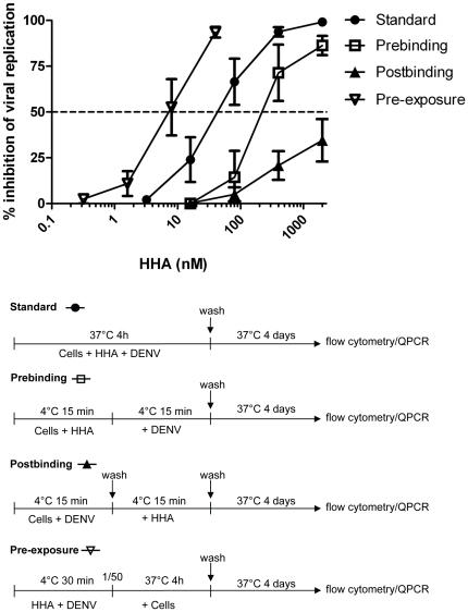
Figure 6. Antiviral assays with DENV-2 in Raji/DC-SIGN+ cells and HHA.
Raji/DC-SIGN+ cells were infected with DENV-2 under four different experimental conditions. (•) Standard antiviral assay: cells were infected with DENV-2 together with HHA (2000-400-80-16-3.2 nM) and incubated at 37°C to allow internalization of the virus. (□) Prebinding assay: cells were pretreated with HHA (2000-400-80-16 nM) before binding to DENV-2. (▴) Postbinding assay: cells were infected with DENV-2 and after washing to remove unbound virus, HHA was added dose-dependently (2000-400-80-16 nM). (∇) Pre-exposure assay: cell-free DENV-2 was pre-exposed to HHA prior to exposure to Raji/DC-SIGN+ cells. Raji/DC-SIGN+ cells were infected with a 50-fold dilution of HHA-exposed DENV (final HHA concentration: 40-8-1.6-0.32 nM). For all four conditions, cells were washed twice to remove virus and/or compound and collected after 4 days. Viral infectivity was analyzed by flow cytometry. Data represent the mean % of inhibition of viral replication ± SEM of 3 independent experiments.

