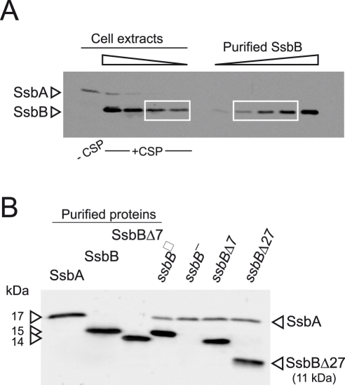
Figure 1. Western blot analysis of S. pneumoniae SsbA and SsbB proteins.
(A) Quantification of SsbB in competent cells. For preparation of cell extracts, 20 ml culture in C+Y medium at OD550 0.13 (∼2.1 108 cfu ml−1) were divided into two equal parts, one of which received CSP (25 ng ml−1), and both were incubated for 12 min at 37°C. Total cells were then collected by centrifugation. Western blotting was as described in Materials and Methods. Volumes of total cell extracts, from left to right: 20 µl (−CSP) and 10, 5, 2.5 and 1.25 µl (+CSP). Amounts of purified SsbB, from left to right: 3.1, 6.3, 12.5, 25 and 50 ng. White rectangles identify cell extract samples and the corresponding purified protein standards used for SsbB quantification. SsbB amounts calculated for 2.5 and 1.25 µl extracts were 30.0 and 14.9 ng, respectively, resulting in an estimate of 70,799±300 SsbB molecules per cell, considering a cell density in the culture of ∼2.7 108 cells ml−1 (because ∼30% of daugther cells remain attached to each other after completion of cell division; our unpublished observations). (B) Western-blot analysis of competent ssbB +, ssbB −, ssbBΔ7 and ssbBΔ27 cells. 5.3 µl of total extracts (corresponding to 235 µl culture at OD550 0.15) were analyzed as described above. Strains used: R1501 (ssbB +), R2294 (ssbB −), R2081 (ssbBΔ7) and R2082 (ssbBΔ27). Purified S. pneumoniae SsbA (50 ng), SsbB (200 ng) and SsbBΔ7 (200 ng) proteins were used as standard.

