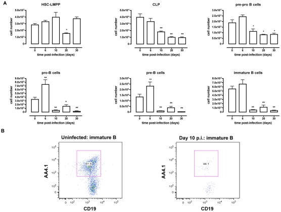
Figure 1. B dyslymphopoiesis in bone marrow during T. brucei infection.
(A) Bone marrow cells from non-infected mice and mice infected with T. brucei for 6–30 days were stained for surface markers commonly used to define developing B cells, as described in table 1 and analyzed using FACS. Data are represented as mean of three mice per group ± SEM, three independent repeat experiments were performed and statistics are compared to uninfected controls (*) p<0,05, (**) p<0,01. (B) Immature B cells were detected as (Lin−B220+IgM+CD43lo/− AA4.1+CD19+) cells in uninfected mice (left panel) versus infected mice on day 10 p.i. (right panel).

