Figure 6.
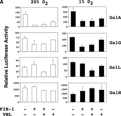
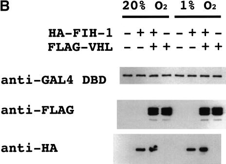
Functional interaction of FIH-1 and VHL to repress HIF-1α transactivation domain function. (A) Hep3B cells were cotransfected with reporters pSV-Renilla and pG5E1bLuc, expression vector encoding the GAL4 DNA-binding domain alone (Gal0) or a GAL4–HIF-1α fusion protein, and expression vectors encoding no protein, FIH-1, or VHL. The GAL4-fusion proteins (containing the indicated HIF-1α residues) tested were GalA (531–826), GalG (757–826), GalL (531–575), and GalH (786–826). The relative luciferase activity represents the ratio of firefly:Renilla luciferase for each construct normalized to the result for Gal0. (B) Immunoblot analysis of lysates from transfected cells using monoclonal antibodies against the GAL4 DNA-binding domain (DBD), FLAG, and HA to detect expression of GalA (top), FLAG–VHL (middle), and HA–FIH-1 (bottom), respectively.


