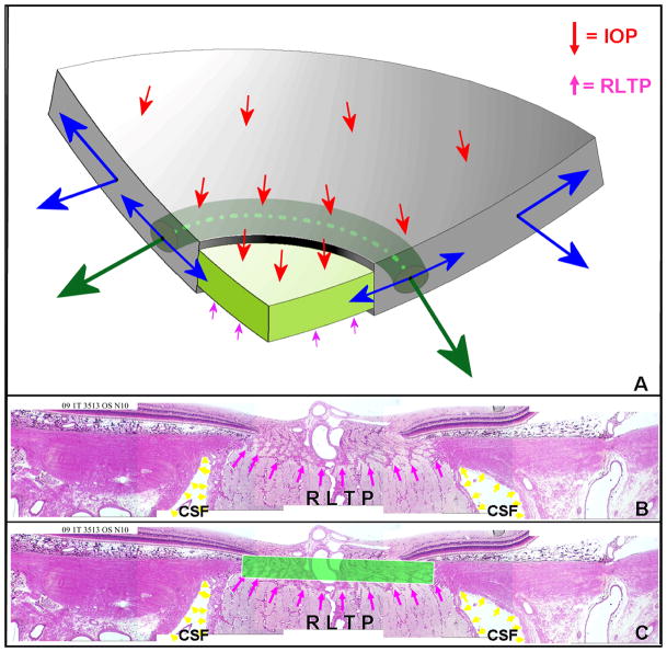
Figure 2. Principle distribution of forces, pressures and the translaminar pressure gradient within the optic nerve head (ONH).
A. Cut-away diagram of intraocular pressure (IOP)-induced mechanical stress in an idealized spherical scleral shell with a circular scleral canal spanned by a more compliant lamina cribrosa. In this case, the majority of the stress generated by IOP/orbital pressure difference (red arrows on the inner surface of the sclera) is transferred into a hoop stress borne within the thickness of the sclera and lamina (blue arrows) that is concentrated circumferentially around the scleral canal (green arrows). B. Note that the pressure behind the lamina is not simply CSF pressure but is retrolaminar tissue pressure (RLTP) which has been demonstrated to be approximately 0.82 × CSF + 2.9 mm Hg by Morgan, et al in dogs (Morgan et al., 1998) C. The difference between IOP and the retrolaminar tissue pressure is the translaminar pressure difference which generates both a net posterior (outward) force on the surface of the lamina (the red arrows over the lamina) and a hydrostatic pressure gradient (the translaminar pressure gradient - schematically shown in green) within the neural and connective tissues of the pre-laminar and laminar regions. Note that the in-plane hoop stress transferred to the lamina from the sclera is much larger than the stresses induced by the translaminar pressure difference. CSF directly influences laminar position through its effect on the translaminar pressure difference. CSF may also effect scleral flange position within the region it projects to the sclera (Figure 2), but in most eyes, because the projection of the CSF space is minimal this is not likely important (the CSF space within Panels B and C in this figure is greatly expanded due to perfusion fixation). IOP has a similar direct effect on laminar position, but has an additional (and potentially more important) effect on laminar position through the peripapillary sclera. However, while the magnitude of the translaminar pressure difference may be small relative to the stresses within the sclera and lamina, the axons experience it as the translaminar pressure gradient the steepness of which is influenced by the thickness of the tissues over which it is experienced. The translaminar pressure gradient, as such, may serve as a primary barrier to axon transport and flow within this region and an important physiologic determinant for the ONH axons and cells. (Panel A is modified from Downs et al, 2009).

