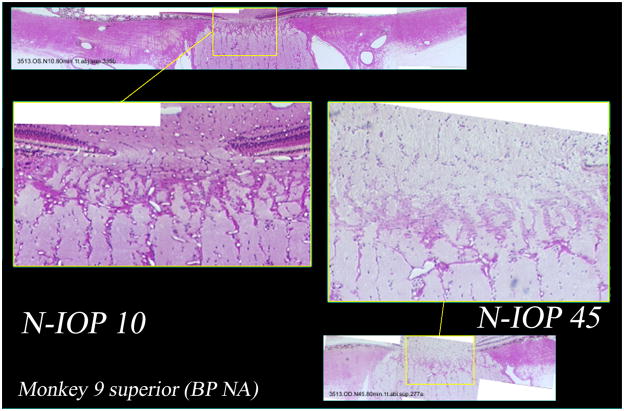
Figure 5. Histologic sections from the superior scleral canal of both eyes of a normal monkey perfusion fixed with one eye at IOP 10 mm Hg (middle left and above) and one eye at IOP 45 mm Hg (middle right and below).
Higher magnification demonstrates patent prelaminar, laminar, and retrolaminar capillaries in the IOP 10 eye (middle left). However in the IOP 45 eye (middle right), the prelaminar and anterior laminar capillaries do not appear patent, with only the posterior laminar and retrolaminar capillaries open.

