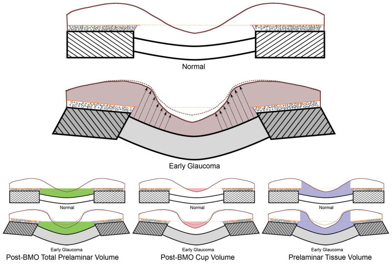
Figure 6. ONH neural and connective tissue changes at the onset of CSLT-detected ONH surface change.
These findings were initially reported in n=3 monkeys (Yang et al., 2007a) and have recently been expanded to n=9 animals (Yang et al., 2010). Normal lamina cribrosa (unhatched), scleral flange (hatched), prelaminar tissue (beneath the internal limiting membrane - brown line), Bruch’s membrane (solid orange line), BMO zero reference plane (dotted orange line), Border tissue of Elschnig (purple line), choroid (black circles) are schematically represented in the upper illustration. Changes in EG are depicted below. We have previously reported the bowing of the lamina and peripapillary scleral flange and thickening of the lamina in these same EG eyes (grey shading) (Yang et al., 2007b). These connective tissue changes underlie posterior deformation of the ONH and peripapillary retinal surface (dotted brown to solid brown ILM) that occurs in the setting of thickening (arrows) not thinning of the prelaminar neural tissues (brown shading) in EG. EG eye expansion of all three volumetric parameters is depicted below. The interactions between these parameters are important. While expansion of the cup and deformation of the surface are clinically detectable at this early stage of the neuropathy, because they occur in the setting of prelaminar tissue thickening, (not thinning), we believe that expansion of Post-BMO Total Prelaminar Volume (due to ONH connective tissue deformation) drives these findings. Thus, cupping in these nine EG eyes is “laminar” in origin, without a significant “prelaminar” component (Yang et al., 2007a).

