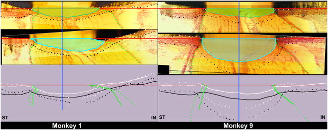
Figure 7. The range of ONH connective tissue deformation (cupping) at the onset of CSLT-detected ONH surface change in nine monkeys with unilateral experimental glaucoma (Yang et al., 2010).
Minimal (Monkey 1, left) to maximum deformation (Monkey 9, right) within the 9 animals is depicted. The following ONH landmarks are delineated within representative superior temporal (ST) to inferior nasal (IN) digital sections from the normal (upper panel) and early experimental glaucoma (EEG) (middle panel) eye of both animals: anterior scleral/laminar surface (white dots), posterior scleral/laminar surface (black dots), neural boundary (green dots), NCO reference plane (red line) and NCO centroid (vertical blue line). Delineated points for the normal (solid lines) and EEG eye of each eye are overlaid relative to the NCO centroid in the lower panel. For each animal, the area under the NCO reference plane and above the anterior laminar surface Post-NCO Total Prelaminar Volume is outlined in both the normal (darker green) and EEG (light blue) eye for qualitative comparison. The overlaid delineations for both eyes of each animal (lower panels) suggest that expansion of this area is due to posterior laminar deformation and neural canal expansion from its onset (Monkey 1) and that these two phenomena continue through more advanced stages of connective tissue deformation and remodeling (Monkey 9). As such,“glaucomatous” deformation of the ONH connective tissues is present early and progresses early in the neuropathy (Burgoyne and Downs, 2008; Yang et al., 2007a). These phenomena are accompanied by regional laminar thickening and outward bowing of the peripapillary sclera in both of these animals and most of the 9 EEG eyes. In addition, in both monkeys laminar migration to the point of partial pialization has occurred on the inferior side (right side of each schematic) of the canal (see Figure 8). (Yang et al., 2010. Optic Nerve Head Lamina Cribrosa Insertion Migration and Pialization in Early Non-Human Primate Experimental Glaucoma. Invest. Ophthalmol. Vis. Sci. 51, ARVO Abstract# 1631)

