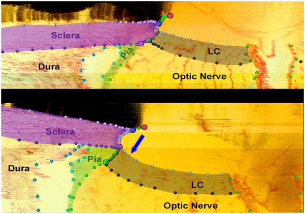
Figure 8. The pathophysiology of early experimental glaucomatous damage to the monkey ONH includes not only “thickening” but regional “migration” of the laminar insertion away from the sclera to the point that “complete pialization” of the laminar insertion is achieved in a subset of eyes.
Neural canal landmarks (Red – Neural Canal Opening (end of Bruch’s Membrane); Blue – Anterior Scleral Canal Opening; Yellow – Anterior Laminar Insertion; Green – Posterior Laminar Insertion; Purple – Posterior Scleral Canal Opening) and segmented connective tissue (dark grey - lamina cribrosa; purple - peripapillary sclera; light green - pial sheath) within digital section images from the inferior region of the normal (top) and the contralateral early experimental glaucoma (bottom) ONH of a representative monkey. Note that in most normal monkey eyes, the lamina inserts into the sclera as is demonstrated in this monkey’s normal eye (top). However at an identical location in the early experimental glaucoma eye of this animal (bottom) in addition to the lamina being thickened and posteriorly deformed, the laminar insertion has migrated outward such that both the anterior and posterior lamina effectively insert into the pial sheath. While regions of laminar insertion into the pia have been reported in normal human eyes (Sigal et al., 2010a), these findings are the first to suggest that active remodeling of the laminar insertion from the sclera into the pia is part of the pathophysiology of “glaucomatous” ONH damage. This phenomenon when present has important implications for the mechanism of axonal insult within these regions. (These data were reported by Hongli Yang, PhD at the 2010 ARVO meeting and are currently in preparation for publication, Yang et al., 2010. Optic Nerve Head Lamina Cribrosa Insertion Migration and Pialization in Early Non-Human Primate Experimental Glaucoma. Invest. Ophthalmol. Vis. Sci. 51, ARVO Abstract# 1631).

