
Figure 1.
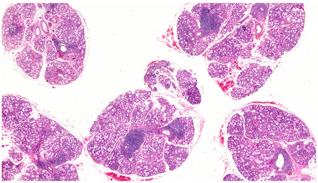
Five hematoxylin and eosin stained labial salivary glands exhibit focal lymphocytic sialadenitis in all glands. About 10 focal lymphocytic infiltrates can be seen in this image. In the microscope, there is a total glandular area of 24 mm2 giving a focus score of 2 foci per 4 mm2. Original magnification ×2.