
Figure 2.
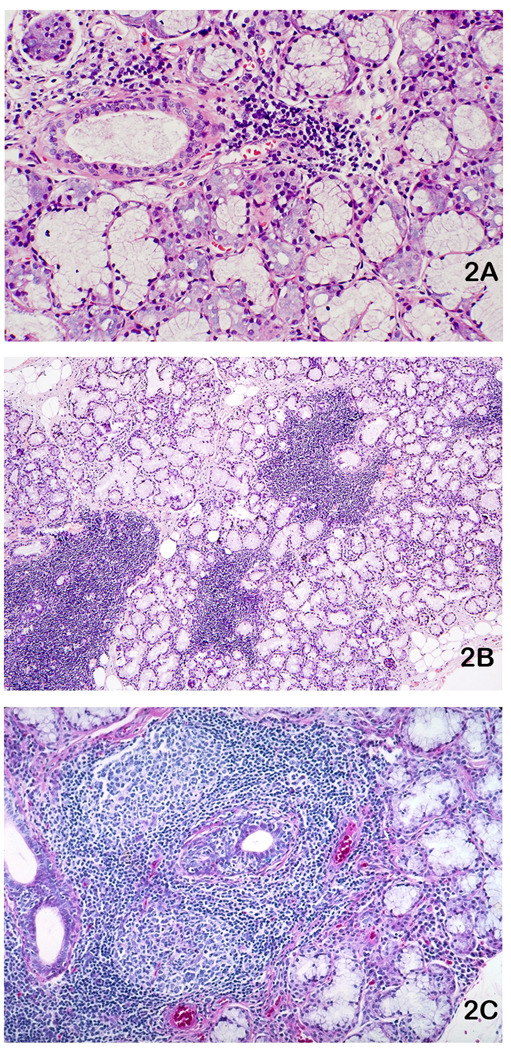
Labial salivary glands (LSG) stained with hematoxylin and eosin exhibiting focal lymphocytic sialadenitis (FLS): A. One LSG with a small lymphocytic aggregate that is minimally sized (> 50 cells) for inclusion in a focus score calculation. Original magnification X100. B. One LSG with four variously sized lymphocytic foci. Note normal appearing acini immediately adjacent to the lymphocyte aggregates, a characteristic feature of FLS. The entire specimen has a focus score of 3 foci per 4 mm2. Original magnification ×16. C. FLS with two prominent lymphocytic germinal centers and ductal hyperplasia with lymphocytic infiltration. Original magnification ×40.