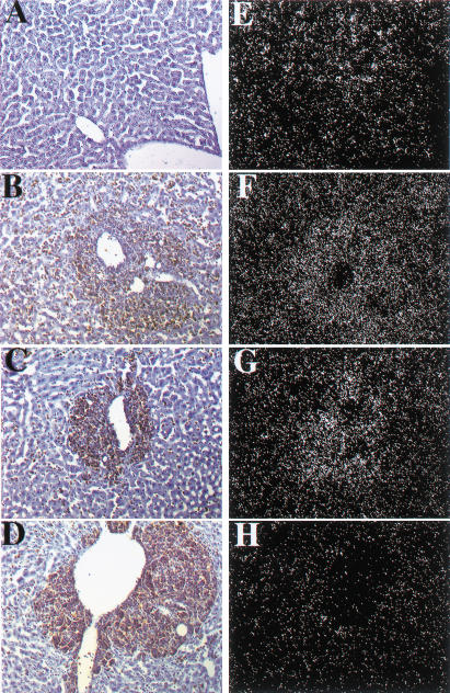
Figure 4.
Detection of Dmp1 RNA by in situ hybridization. Liver cells from Dmp1+/+ mice (A), stained with hematoxylin (blue) express Dmp1 RNA (E). Malignant lymphoma cells in Eμ-Myc transgenic mice, visualized by antibody staining for B220 antigen (brown), surrounded central veins and invaded adjacent liver sinusoids (B–D). Regions containing metastatic B-cells in livers from both Eμ-Myc Dmp1+/+ (B) and Eμ-Myc Dmp1+/− (C) mice revealed increased hybridization signals with a Dmp1 antisense probe (F,G), respectively, relative to that in normal liver (E), consistent with continued Dmp1 expression in lymphomas from heterozygous mice. Similar results were obtained in three of three Dmp1+/− animals. Background hybridization with tissues from Eμ-Myc Dmp1−/− mice (H) was equivalent to that obtained with a control Dmp1 sense probe (data not shown).

