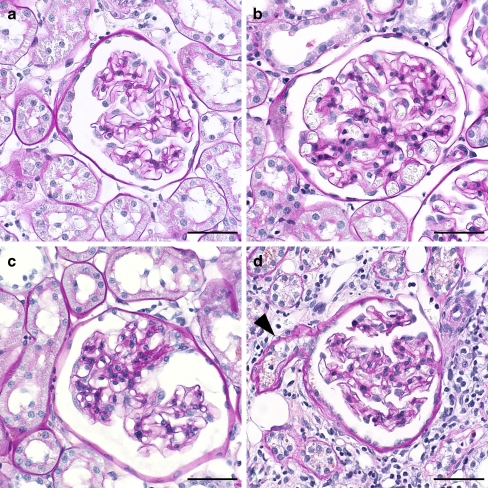
Fig. 1.
Representative glomeruli from the four groups. a Normal glomerulus from WKY rat with segmental thickening and lamellation of Bowman’s capsule. b Normal glomerulus from SHR. c Glomerulus with segmental sclerosis from SHR. d Glomerulus from SHR with collapsed capillary convolute and thickened and lamellated Bowman’s capsule. The outlet of the atrophic proximal tubule is also seen (arrowhead). (All images scale bar 50 μm, PAS stain)

