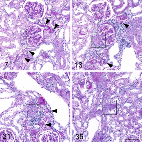
Fig. 2.
Example from serial sections of a nephron with glomerular capillary collapse in a SHR. Numbers indicate the consecutive numbers of the serial sections beginning with the number 1 for the first section of the glomerulus. Arrowheads point to related proximal tubules. Rectangles indicate afferent arteriole, the circle efferent arteriole. The glomerulus shows focally thickened Bowman’s capsule, there are no signs of segmental sclerosis in the collapsed tuft. The proximal tubule is atrophic with reduced diameter and thickened basement membrane. The tubular neck area is seen in image 7. The wall of the afferent arteriole is hypertrophic. Image 35 shows the outlet of the afferent arteriole from the interlobular artery. (All images PAS stain, scale bar 100 μm)

