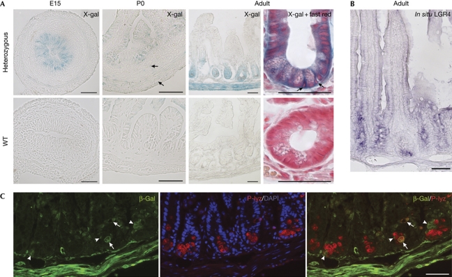
Figure 1.
Lgr4 expression pattern in the ileum. (A) Lgr4/LacZ expression detected by X-gal staining of heterozygous or wild-type (WT) embryonic (E15), newborn (P0) and adult mice. Arrows in newborn heterozygous panel indicate faint but specific X-gal-positive mesenchymal or smooth muscle cells. In adults, arrows indicate CBC cells in an enlarged view of X-gal-stained crypt with fast red counterstaining. (B) In situ hybridization of an adult section showing Lgr4 expression all along the crypt. (C) Co-staining of β-gal and P-lyz antibodies with DAPI. In the bottom of the crypts, β-gal-expressing cells that do not express the P-lyz Paneth cell marker are shown (arrowheads), whereas few cells show double staining (arrows). Scale bars, 50 μm. β-gal, β-galactosidase; DAPI, 4,6-diamidino-2-phenylindole; E, embryonic day; P, postnatal day; P-lyz, P-lyzozome; WT, wild type; X-gal, 5-bromo-4-chloro-3-indolyl-D-galactoside.

