Abstract
The authors report a case of atherosclerotic stroke in a 46-year-old recreational bodybuilder with a 20 year history of anabolic-adrenergic steroid (AAS) abuse. Cerebrovascular accident (CVA) occurred during his third week of hospital admission for an acute abdomen and on day 8, postemergency laparotomy. CVA presented with collapse, generalised seizures, reduced Glasgow Coma Score and severe hypertension. He was subsequently admitted to the intensive care unit (ICU), where initial investigations did not illustrate an underlying diagnosis. By day 4 in ICU, there had been no significant clinical improvement and radiological investigations were repeated, identifying a left frontal lobe infarct in the middle cerebral artery territory. The authors propose CVA was secondary to AAS. After a prolonged and complicated period of rehabilitation, he has been discharged home; he requires carers due to dyspraxia and is mobilising independently.
Background
Anabolic-adrenergic steroids (AAS) are commonly being self-administered in supraphysiological doses by recreational bodybuilders to increase muscle mass, strength and enhance athletic performance. There are numerous multi-organ side effects of AAS abuse (table 1), many of which are dose-related and reversible upon drug cessation.1
Table 1.
Side effects of AAS7
| Acne vulgaris | Psychiatric: depression, psychosis |
| Dyslipidaemia | Glucose intolerance |
| Cholestatic jaundice | Hepatocellular carcinoma |
| Oligo-azospermia | Painful gynaecomastia |
| Musculoskeletal: tendon injuries | Renal carcinoma |
The authors report a case of stroke in a recreational bodybuilder with a history of nandrolone abuse and drug related co-morbidities: obstructive sleep apnoea (OSA), chronic renal failure, osteoarthritis and depression with 3 prior suicide attempts.
Cerebrovascular accidents (CVA) are a rare, but significant risk of AAS and few reported cases of ischaemic stroke in AAS users exist.2–5 This case is the first, to our knowledge of ischaemic stroke in a nandrolone user. It highlights the importance of recognition of AAS abuse in patients and knowledge of the consequences.
Case presentation
The patient, a 46-year-old recreational bodybuilder, had a 20 year history of nandrolone abuse, alongside intermittent use of growth hormone and testosterone. He injected Deca-Durabolin, a form of nandrolone and would administer this in increasing doses over a 2–3 month period and follow this ‘buildup cycle’ with a drug-free period. This patient’s average dose of nandrolone varied enormously, while the cyclical administration pattern remained continuous. At hospital admission, he weighed 134 kilograms and had a well defined muscular physique.
Our patient was admitted to hospital twice within a week; the first admission was for intravenous antibiotic treatment of a lower limb cellulitis. He was discharged home after 5 days and was readmitted 1 day later with abdominal pain and severe sepsis. CT abdomen showed acute cholecystitis and blood cultures were positive for Escherichia coli. On the surgical ward, pain and pyrexia persisted despite treatment and he was re-imaged 11 days later, when a perforated sigmoid diverticulum was identified. He was subsequently admitted to the intensive care unit (ICU) following an emergency laparotomy. Pain control issues continued postoperatively and high doses of morphine through a patient controlled analgesia device were required to keep him comfortable. Hypertension was an intermittent problem and it was felt that addressing his pain was the optimum way to control his blood pressure. Otherwise, he continued to make a good recovery and was discharged to the surgical ward.
Three days later, he was readmitted to ICU following a collapse with witnessed generalised seizures, a Glasgow Coma Score of 3/15, hypoxia, tachycardia, severe hypertension and temperature of 38°C. He required immediate intubation and ventilation for oxygenation.
Significant findings of initial investigations showed a respiratory acidosis on blood gas analysis, white blood cell count 44.3, C reactive protein 146 and troponin-T 1.3 with a normal ECG and 24 h tape. Radiological investigations included a normal CT head scan, a CT abdomen showing changes consistent with recent surgery and a CT pulmonary angiogram that showed features in keeping with acute respiratory distress syndrome. He was subsequently ventilated using high frequency oscillation for 12 h and required 2 days of inotrope support.
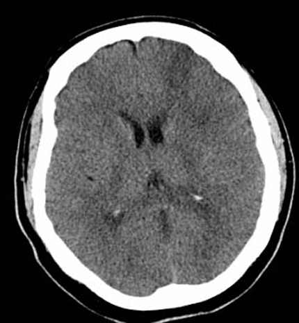
Intermittent right-sided seizures persisted for 2 days despite phenytoin therapy and were attributed to sepsis and hypoxia. Sedation was turned off on day 4 and a neurological examination showed him to have right-sided upper limb weakness (Medical Research Council (MRC) scale 0/5), that was greater than his lower limb (MRC scale 4/5), with hypertonia, an extensor plantar response and right-sided neglect. There was no response to antibiotics and a repeat CT head scan revealed an extensive left frontal lobe infarct in the middle cerebral artery (MCA) territory (figure 1).
Figure 1.

CT head showing left middle cerebral artery infarct.
Investigations
Further investigations shed no light on the cause of CVA and included a carotid artery duplex scan, a transoesophageal echo with bubble study, auto-antibody, lupus and vasculitis screen. However, a lipid profile revealed total cholesterol of 3.4 mmol/l (normal value <5 mmol/l) and a raised low-density lipoprotein (LDL) cholesterol of 2.7 mmol/l (normal value <2.0 mmol/l).
Differential diagnosis
Prior to obtaining the repeat CT head scan, underlying differential diagnoses included meningitis, encephalitis, central line, chest or abdominal sepsis.
Outcome and follow-up
After a prolonged stay on the stroke unit with extensive rehabilitation, our patient unfortunately encountered further complications suffering a pulmonary embolism that is being managed with lifelong warfarin in view of his second thromboembolic event and pancreatitis, which was managed conservatively. He has now been discharged home, where he is independently mobilising, but has residual right upper limb weakness. His main problem is dyspraxia and he requires carers to assist with functional daily tasks.
Discussion
The link between stanozolol and CVA has been recognised, but to date there have been no case reports of nandrolone and CVA. Nandrolone is the most commonly used injectable steroid and is a 17-β-ester testosterone derivative, while stanozolol is a 17-α-alkyl testosterone derivative and can be administered orally or intramuscularly.6 Risk of CVA is enhanced with concurrent use of other doping substances.7 There have been no documented cases of CVA in AAS users that have been concurrently administering growth hormone or testosterone. Our patient had not used growth hormone or testosterone within the year prior to his CVA.
On admission, medical history included OSA, chronic renal failure, osteoarthritis and depression. While these co-morbidities are significant, none are known to have causal links with CVA in this patient’s age group. OSA, osteoarthritis and depression have been associated with AAS and it is likely they are secondary to nandrolone use in this patient.7
Proposed mechanisms of AAS induced CVA includes atherosclerosis, hyperhomocysteinemia, vasospasm and platelet aggregation; complex interactions exist between these four mechanisms.
Atherosclerosis occurs secondary to decreased high-density lipoprotein and increased LDL cholesterol in the presence of normal total cholesterol and triglyceride levels.6 Dyslipidemia results in endothelial dysfunction, increased risk of atheroma formation and subsequently increased risk of CVA. Our patient’s lipid profile illustrates a pattern consistent with AAS use.
Increased homocysteine levels also impair endothelial function. AAS users have significantly elevated levels of homocysteine,8 which rise most significantly during the higher dose phase of the AAS cycle.6 Graham et al, 2006 proposes homocysteine induces endothelial dysfunction by generation of reactive oxygen species and the use of antioxidants by AAS users may be beneficial in modifying these effects.
Arterial vasospasm occurs due to excess LDL cholesterol oxidising at the endothelium and inhibiting endothelial nitric oxide release. Decreased nitric oxide production impairs smooth muscle relaxation and vasodilatation.2 This is a similar pathophysiological mechanism to that seen in cocaine and nitric oxide, which is known to induce coronary artery vasospasm. Cocaine also affects other parts of the circulation as illustrated by ischaemic damage to the nasal septum. As the endothelial effects of nitric oxide are similar throughout the vascular tree, such as glyceryl trinitrate induced headache, it seems likely AAS may cause cerebral artery vasospasm.
Effects on the haemostatic system increase the risk of thrombosis formation. Decreased nitric oxide increases endothelial production of thromboxane A2 and decreases production of prostacyclin. This causes enhanced platelet sensitivity and aggregation, despite a normal platelet count,2 8 and therefore increased CVA risk.
On a more positive note, AAS and in particular nandrolone, may have some beneficial effects when administered in low doses under medical supervision in the management of isolated postoperative muscle wasting and generalised muscle wasting after periods of immobilisation.
Greater knowledge and appreciation of AAS would have altered our patient’s management and may have even prevented the CVA from occurring. Firstly, exploration of his drug history and links with his co-morbidities did not take place until his initial admission to ICU. This should have been explored on admission to hospital and resulted in increased recognition of the severity of the patient’s condition and prompted earlier investigation. Secondly, more aggressive management of his hypertension should have occurred throughout hospital admission. Thirdly, an electroencephalograph should have been performed following his collapse, as this may have revealed an ischaemic focus prior to any radiological changes. Finally, with the above knowledge, CVA as the unifying diagnosis should have been considered earlier as a cause of collapse and thrombolysis administered despite relative contraindications.
This case is the first to demonstrate a link between nandrolone and CVA and adds to the mounting evidence in the literature that cerebrovascular side effects of AAS must be recognised. We have proposed four mechanisms linking AAS and cerebrovascular morbidity; however, the pathogenesis is multifactorial and there is significant overlap between each mechanism. It is clear that further research is required to clarify the thrombogenicity of AAS and to generate direct evidence of the effects on humans.
Learning points.
-
▶
Have a high index of suspicion for AAS abuse and obtain detailed drug histories in a general practice and hospital setting.
-
▶
Be aware of and recognise the multiple co-morbidities associated with AAS use.
-
▶
When AAS abuse is known, actively look for other co-morbidities and try to identify and modify risks.
Footnotes
Competing interests None.
Patient consent Obtained.
References
- 1.Hartgens F, Kuipers H. Effects of androgenic-anabolic steroids in athletes. Sports Med 2004;34:513–54 [DOI] [PubMed] [Google Scholar]
- 2.Santamarina RD, Besocke AG, Romano LM, et al. Ischemic stroke related to anabolic abuse. Clin Neuropharmacol 2008;31:80–5 [DOI] [PubMed] [Google Scholar]
- 3.Frankle MA, Eichberg R, Zachariah SB. Anabolic androgenic steroids and a stroke in an athlete: case report. Arch Phys Med Rehabil 1988;69:632–3 [PubMed] [Google Scholar]
- 4.Akhter J, Hyder S, Ahmed M. Cerebrovascular accident associated with anabolic steroid use in a young man. Neurology 1994;44:2405–6 [DOI] [PubMed] [Google Scholar]
- 5.Laroche GP. Steroid anabolic drugs and arterial complications in an athlete–a case history. Angiology 1990;41:964–9 [DOI] [PubMed] [Google Scholar]
- 6.Kam PC, Yarrow M. Anabolic steroid abuse: physiological and anaesthetic considerations. Anaesthesia 2005;60:685–92 [DOI] [PubMed] [Google Scholar]
- 7.Quaglio G, Fornasiero A, Mezzelani P, et al. Anabolic steroids: dependence and complications of chronic use. Intern Emerg Med 2009;4:289–96 [DOI] [PubMed] [Google Scholar]
- 8.Graham MR, Grace FM, Boobier W, et al. Homocysteine induced cardiovascular events: a consequence of long term anabolic-androgenic steroid (AAS) abuse. Br J Sports Med 2006;40:644–8 [DOI] [PMC free article] [PubMed] [Google Scholar]


