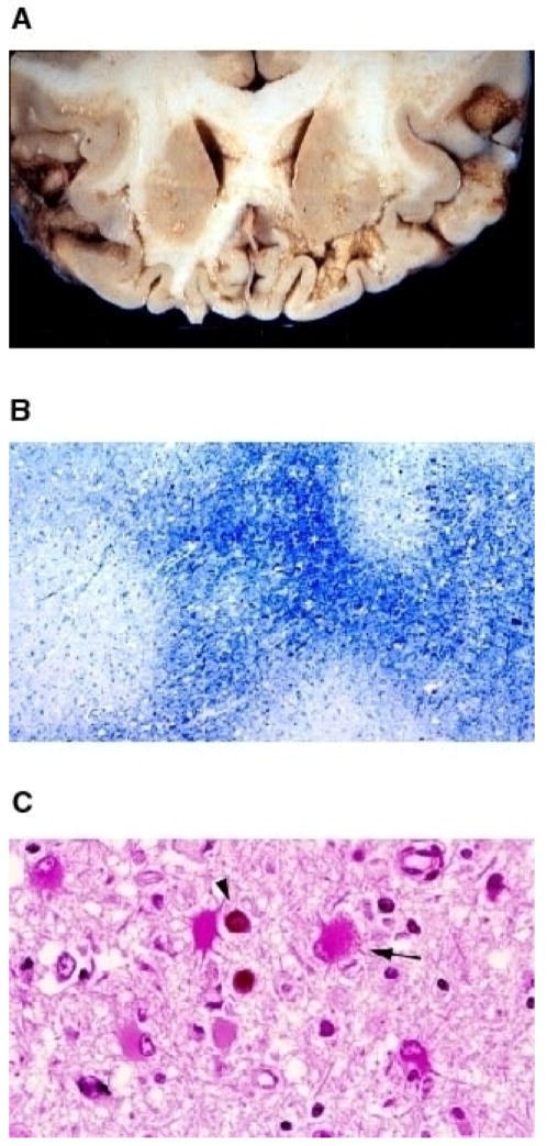
Fig. 1. Histological features of PML.

(A) Gross examination of JCV- induced lesions occurring at the subcortical white matter. A coronal section of the frontal lobe of the brain from a PML patient is shown. (B) Apparent myelin loss, as result of JCV infection of oligodendrocytes, is made detectable by Luxol blue staining (40×). Demyelinated areas are visibly distinguishable as white plaque areas. (C) Hematoxilin and Eosin staining of the brain sections from a PML patient. Infected oligodendrocytes are indicated with a round dark staining of the eosinophilic inclusion bodies (arrow head). An arrow points to an infected astrocyte (400×). Reproduced with permission from (111).
