Figure 5.
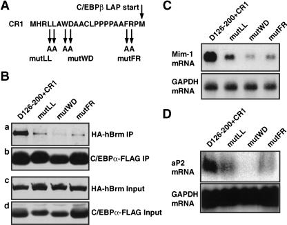
Mutation analysis of CR1. (A) Amino acid sequence of mouse CR1, indicating the positions of residues mutated to alanines in the mutLL, mutWD, and mutFR CR1 variants, respectively. (B) Coimmunoprecipitation analysis of C/EBPα D126–200+CR1 and its mutant derivatives was performed as in Figure 2B. The HA–hBrm coprecipitated with the C/EBPα–FLAG-derivatives is shown in panel a, the precipitated C/EBPα–FLAG in panel b. The levels of the same proteins in the input lysate for the coimmunoprecipitation are shown in panels c and d, respectively. (C) The ability of C/EBPα D126–200+CR1 and its mutant derivatives to induce mim-1 expression in HD3 erythroblasts was analyzed as in Figure 3B. (D) The induction of aP2 mRNA in retrovirally transduced NIH3T3 cells determined as in Figure 1B.

