Fig. 4.
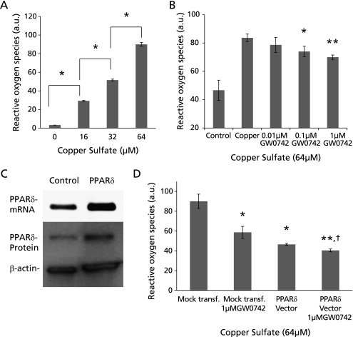
Copper-induced reactive oxygen species formation in HepG2 cells. (A) Copper at concentrations of 16, 32 and 64 µM induced ROS formation in a dose dependent manner (*p<0.0005). (B) ROS induced by 64 µM of copper were significantly lower in HepG2 cells co-treated with 0.1 and 1 µM of GW0742 (*p<0.05, **p<0.01 vs copper). (C) Transfection of HepG2 cells with our PPARδ expression vector effectively increases PPARδ mRNA and protein. (D) HepG2 cells transfected with PPARδ expression vector and exposed to 64 µM of copper had significantly less ROS than those transfected with an empty vector and exposed to 64 µM of. Cells transfected with PPARδ expression vector and treated with 1 µM GW0742 had an additive effect decreasing copper-induced ROS formation copper (*p<0.05, **p<0.01 vs mock transfection; †p<0.05 vs PPARδ vector). Error bars represent SEM and p value was assessed by Student’s t test.

