Fig. 2.
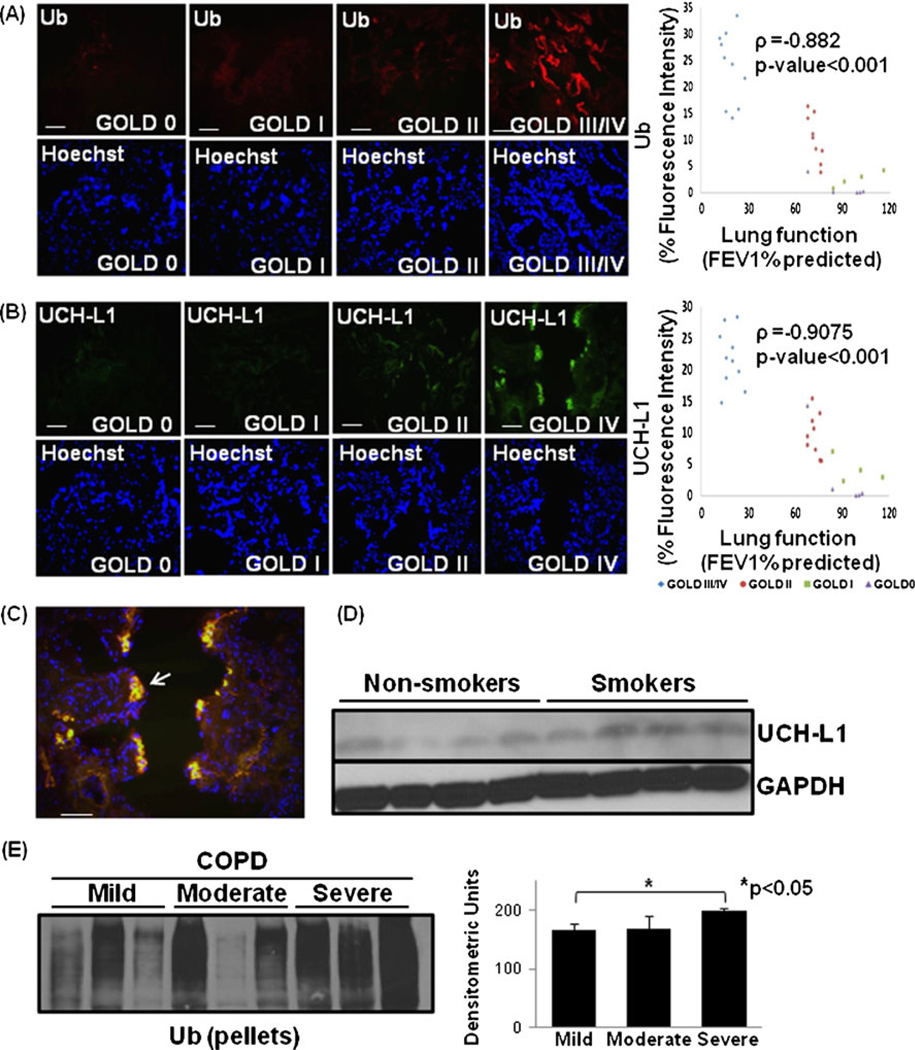
Accumulation of ubiquitinated proteins in severe emphysema. a The paraffin-embedded longitudinal human lung sections of control (GOLD 0) and COPD lung tissues at GOLD I–IV levels of emphysema (n=8–10, each group), immunostained with primary mouse monoclonal Ub (P4D1) and secondary anti-mouse-FITC conjugated antibody, present significant increase in protein levels and accumulation of ubiquitinated proteins in the human lung tissue sections with severe (GOLD III/IV) emphysema. b Lung sections (from a) immunostained with primary rabbit polyclonal UCH-L1 and secondary anti-rabbit-FITC conjugated antibody, show significant increase in its expression and accumulation with increasing severity of emphysema. Nuclear (Hoechst) staining of the same area is shown in the bottom panel. Scale=50 µm. Densitometric analysis confirms association of Ub and UCH-L1 (p<0.001) with progressive lung inflammation and severity of emphysema. c The data show the co-localization of ubiquitin and UCH-L1 in severe COPD lung tissue sections. Scale=50 µm. d This is further confirmed by the up regulation of UCH-L1 in smokers as compared with normal controls. The GAPDH shows the equal loading. e Immunoblot analysis of protein pellets of lung tissues from COPD smokers with mild, moderate and severe emphysema verify the higher accumulation of poly-ubiquitinated insoluble protein aggregates in severe emphysema (p<0.05). The data confirm the accumulation of poly-ubiquitinated proteins in insoluble protein fractions of COPD lungs as a potential mechanism for pathogenesis of severe emphysema

